January 27, 2025
Diagnosis (Ocular)
How is Ocular Melanoma Diagnosed?
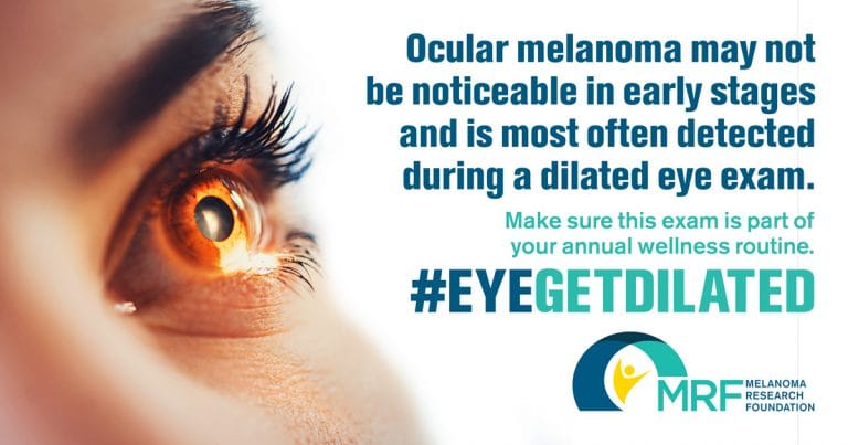
Ocular melanoma (OM) is most often detected by an optometrist or an ophthalmologist during a dilated eye exam. OM is frequently asymptomatic until the tumor grows large enough to create visual changes, such as blurred or distorted vision, flashes, or loss of peripheral vision. Only iris melanoma can sometimes be detected by external examination (looking at the outside of the eye).
After an OM diagnosis, your doctor may order imaging tests such as an ultrasound of the eye to determine the size of the tumor, and scans like MRI, CT, or PET to check for signs of spread beyond the eye.
Unlike other forms of melanoma, a biopsy is not usually performed to diagnose OM. Instead, OM is considered a clinical diagnosis – meaning it is typically made based on the appearance of the tumor during an eye exam and imaging results.
If you or a loved one have been diagnosed with OM, or to learn more about OM research, treatment and support resources, please visit the MRF’s CURE Ocular Melanoma (CURE OM) initiative.

Introducing the New Ocular Melanoma Patient Guide!
Genetic Testing, Tumor Size and Metastatic Risk
Once ocular melanoma (OM) is diagnosed, several factors should be discussed with your treatment team to better understand your specific diagnosis. While treating the primary eye tumor remains the most important initial step, assessing the risk of metastatic disease (spread to the other parts of the body) is also a key part of care. Factors associated with risk include tumor size and location, and genetic features.
Common Genetic Tests in OM
Genetic features can provide important insight into metastatic risk. This information is obtained through a biopsy, where tumor cells are tested for changes in certain genes or chromosomes. The results of these tests can help your treatment team develop an appropriate and individualized surveillance plan and, if necessary, a treatment plan.
Timing is critical because:The biopsy must be performed before the tumor is treated with radiation, as radiation can damage DNA and affect test results.
Two different types of genetic testing may be performed:
1) CHROMOSOME ANALYSIS (KARYOTYPING)
Changes in certain chromosomes (the packages of DNA inside cells) can show higher risk. The most important are:
- Loss of chromosome 3 (called monosomy 3) — linked to a higher chance of metastasis
- Gain of part of chromosome 8 (called 8q gain) — also linked to higher risk
- Changes in chromosomes 1 and 6 may be seen too, but are less reliable indicators.
2) GENETIC EXPRESSION PROFILE (GEP) TESTING
This test looks at the “molecular signature” of the tumor using a 15-gene panel. Based on the results, tumors are grouped into risk classes:
- Class 1A tumors have a low risk of metastasis
- Class 1B tumors have an intermediate risk of metastasis
Class 2 tumors have a high risk of metastasis
Should I have my tumor tested?
Many patients say they prefer to know their risk if given the choice. Even if the results are worrisome, knowing can help patients and doctors plan follow-up care more closely and sometimes detect spread earlier. Deciding whether to do testing is personal, and it may help to talk with your doctor or a genetic counselor about timing, insurance coverage, and what the results could mean for you.
Genetic Mutations in Ocular Melanoma
A variety of genetic mutations have been found in OM. These mutations are thought to “drive” the disease:
- GNAQ and GNA11 – GNAQ and GNA11 mutations are the most common mutations in uveal melanoma, appearing in more than 80% of all cases. These mutations do not seem to be associated with patient outcomes or risk of metastasis.
- BAP1 – BAP1 mutations are found in about half of uveal melanoma cases and are associated with a high risk for metastasis. The BAP1 mutation is also strongly associated with a Class 2 gene expression profile (GEP).
- SF3B1 – SF3B1 mutations (seen in ~ 10-20% of cases) are often linked with intermediate risk of metastasis
- EIP1AX — EIF1AX mutations (seen in ~10%) are generally linked with a better prognosis and lower risk of metastasis.
- BRAF – BRAF mutation are common in cutaneous melanoma but are rare in ocular melanoma. They can be found in conjunctival melanoma cases.
Speak with your oncologist about what your mutation status could mean and when your tumor should be tested.
What does this mean for treatment?
At this time, these risk factors do not change standard treatment for the primary eye tumor. However, they can help guide how closely you are monitored after treatment and whether you might be eligible for a clinical trial. Ongoing studies are exploring treatments given before or after eye-directed therapy to reduce the risk of spread, and your doctor can let you know if any are appropriate for you.


