

March 31, 2026

Melanoma Education
Melanoma doesn't discriminate.
What Is Skin of Color?
According to the Skin of Color Society, “People with skin of color are of diverse racial and ethnic backgrounds, and include African Americans, Asians, Hispanics or Latinos, Native Indians and Pacific Islanders primarily.”

Although melanoma is relatively uncommon in people of color, it is often diagnosed at more advanced stages-making it more difficult to treat and more likely to be fatal. Studies consistently show that people of color are more likely to die from skin cancer compared to whites.¹

Studies have shown that the 5-year survival rate for melanoma is lower in people of color compared to White individuals, with Black patients having a survival rate of approximately 67% compared to 92% in White patients.¹ These gaps have persisted even as advances in treatment have improved outcomes overall, which is why early detection is so important.

Studies reveal that people of color receive little or no skin education from their doctors about the risk and prevention of skin cancer.⁴
While UV exposure plays less of a role in the development of melanoma in people of color, it often plays a less dominant role compares to lighter skin types, with other contributing risk factors including:
Impaired-Immune System
Prior Radiation Therapy
Preexisting Pigmented Lesions
Albinism
Chronic Scars Burn Scars
Effects of Melanoma on Skin of Color.
In non-Hispanic White individuals, more than 90% of cutaneous (skin) melanomas are thought to be linked to UV exposure.6 This includes UV rays that come from either natural sunlight or from artificial sources like tanning beds or sun lamps.
In people of color, melanomas most commonly develop on areas that receive little to no sun exposure, such as the palms of the hands, soles of the feet, under the nails and mucosal sites like the mouth, nasal passages or genitals, rather than on sun-exposed skin.8 This is different from melanoma in non-Hispanic White individuals, where UV exposure is the primary driver, Melanomas developing in these locations may be classified as acral lentiginous melanomas or mucosal melanoma. Melanoma can also occur in the eye and is known as ocular melanoma.

Facts & Stats for Melanoma Skin of Color in the U.S.²
Melanoma incidence rates vary by racial group.

1 PER 100,000 MEN: 0.9 PER 100,000 WOMEN
African-American

1.36 PER 100,000
Asian/Pacific Islander

4.9 PER 100,000
Hispanic

10.7 PER 100,000 (13.0 MEN; 9.2 WOMEN, AND RISING)
Alaskan/Native Indian

34.7 PER 100,000 (34.7 MEN; 22.1 WOMEN)
Non-Hispanic White
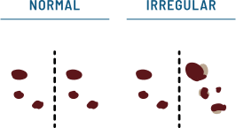
What Does Melanoma Look Like?
Having your skin checked once each year by a dermatologist, as well as checking your skin at home each month, may help melanoma be caught in its earliest stage. Although melanoma can only be diagnosed with a biopsy, the ABCDE rule can help you and your dermatologist identify a melanoma. Note that melanoma in people of color may not always follow the classic ABCDE criteria. It may appear symmetric with uniform pigmentation, or present as a hypomelanotic (lightly pigmented) lesion, making it especially important to have all suspicious lesions evaluated by a dermatologist.
A

Asymmetrical Shape
Melanomas are often irregular, or not symmetrical, in shape. Benign moles are usually symmetrical.
B

Border
Typically, non-cancerous moles have smooth, even borders. Melanomas usually have irregular borders that are difficult to define.
C

Color
The presence of more than one color (blue, black, brown, tan, etc.) or the uneven distribution of color can sometimes be a warning sign of melanoma. Benign moles are usually a single shade of brown or tan.
D

Diameter
Melanomas are often greater than 6 millimeters in diameter (approximately the size of a pencil eraser).
E
Evolution (or CHANGE)
The evolution of your mole(s) has become the most important factor to consider when it comes to diagnosing a melanoma. Knowing what is normal for YOU could save your life. If a mole has gone through recent changes in color and/or size, bring it to the attention of a dermatologist right away.
How Can You Detect Skin Cancer Early?
The key is early detection, and it starts with you. The best way to find skin cancer is to check your own skin.
This is crucial because when performed monthly, you can find changes to spots in your skin and get treated appropriately. When detected early, skin cancer can be treated and often cured. However, in later stages, skin cancer can turn deadly, making treatment difficult.

Here's What Dermatologists Recommend
For People Who Have Skin of Color

Self Exam
Full body exam of your skin

Use
Full-length mirror and a partner or a handheld mirror

Do
Monthly
Things to look for:
Sores that won't heal or heals and then returns
Sore that has a hard time healing. Pay special attention if the sore appears in a scar or on skin that was previously injured.
Patch or spot on skin that feels rough and dry
Dark line or spot underneath or around a fingernail, toenail, or palms of hands and feet
Dark spot, growth, or darker patch of skin that is growing, bleeding or changing in any way
For lesions on the feet and nails, use the CUBED criteria: Colored, Uncertain, Bleeding, Enlarged, Delayed healing. Any of these features warrants evaluation.
Where to Look:
Look at your skin from head to toe
Examine the top of your head and back using a handheld mirror
Check places that get minimum exposure — bottoms of your feet, toenails, lower legs, groin, buttocks, inside of your mouth, around and underneath your fingernails and mucosal surfaces (gums, palate, genitals)
If you find something, consult a dermatologist
To learn more about how to conduct a self-screen exam.
Things you can do to protect yourself.

Stay in the shade whenever possible.

Wear sun-protective clothing.

Stay away from tanning beds.
Wear sunscreen daily.
Why Wearing Sunscreen Daily Is So Important.
- Dermatologists recommend: SPF 30+, broad-spectrum sunscreen, and water resistant.
- Apply sunscreen 15-30 minutes before going outside.
- Reapply every 2 hours and more regularly if you’re sweating or getting wet.

Citations:
Updated 2026
Qian Y, Johannet P, Sawyers A, Yu J, Osman I, Zhong J. The ongoing racial disparities in melanoma: an analysis of the Surveillance, Epidemiology, and End Results database (1975–2016). J Am Acad Dermatol. 2021;84(6):1585-1593. doi:10.1016/j.jaad.2020.08.097
Gohara MA. Skin cancer in skins of color. J Drugs Dermatol. 2008;7:441–45
Pipitone M, Robinson JK, Camara C, Chittineni B, Fisher SG. Skin cancer awareness in suburban employees: A Hispanic perspective. J Am Acad Dermatol. 2002;47(1):118-123. doi:10.1067/mjd.2002.120450
Kim M, Boone SL, West DP, Rademaker AW, Liu D, Kundu RV. Perception of skin cancer risk by those with ethnic skin. Arch Dermatol. 2009;145(2). doi:10.1001/archdermatol.2008.566
Lucas RM, McMichael AJ, Armstrong BK, Smith WT. Estimating the global disease burden due to ultraviolet radiation exposure. Int J Epidemiol. 2008;37(3):654-667
Gloster HM, Neal K. Skin cancer in skin of color. J Am Acad Dermatol. 2006;55:741-60
Bradford PT, Goldstein AM, McMaster ML, Tucker MA. Acral lentiginous melanoma: incidence and survival patterns in the United States, 1986-2005. Arch Dermatol. 2009;145(4):427-434. doi:10.1001/archdermatol.2008.609
Brunsgaard EK, Wu YP, Grossman D. Melanoma in skin of color: Part I. J Am Acad Dermatol. 2023;89(3):445-456. doi:10.1016/j.jaad.2022.04.056
Townsend JS, Melkonian SC, Jim MA, Holman DM, Buffalo M, Julian AK. Melanoma incidence rates among non-Hispanic American Indian/Alaska Native individuals, 1999–2019. JAMA Dermatol. 2024;160(2):148-155. doi:10.1001/jamadermatol.2023.5226